Fetal Brain Doc
Fetal brain outpatient care
*All patients who undergo mid-term fetal Dock and late stage fetal Dock at CRIFM include a Fetal Brain Dock.
For mothers who are worried about abnormal findings in the brain
Some mothers may have concerns about the development of their fetus after a prenatal checkup, such as the size of the ventricles or shadows seen on the brain.
Fetal brain outpatient care can take images of the brain by ultrasound and check the developmental status against the number of weeks.
- If you have been told that your fetus has a large or small head
- If you have been told that your fetus has large ventricles or hydrocephalus
- You have been told that your unborn child has something in the brain.
- Your older child has a developmental disability.
- If your older child has been treated for hydrocephalus, brain disease, or skull disease.
- Your older child has a disease caused by genetic mutation such as Joubert syndrome, hereditary hydrocephalus (L1CAM mutation), Lissencephaly, etc.
- Those who are concerned about their baby's brain development due to medication or viral infection during pregnancy.
In any patient, the fetal Dock of CRIFM is to see if the brain is developing properly in that number of weeks.
What we know about Fetal brain outpatient care
The fetal brain changes its shape significantly with each passing week. It is very important that the brain is well developed at each week. Recently, it has become possible to predict cases of poor development of the cerebral cortex as early as 18 to 20 weeks of pregnancy.
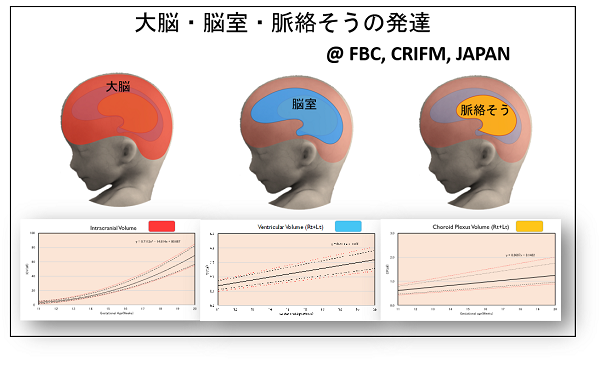
In CRIFM, a special ultrasound called fetal neurosonography is used to depict the brain of a fetus more clearly than MRI, to measure the angle and size of fine brain structures in three dimensions, and to check the growth of blood vessels.
There are many reasons to be concerned about the brain development of a fetus, such as an enlarged ventricle, a shadow on the brain, a suspicion of hydrocephalus, an older child with a brain disease, or a hereditary disease. There are also cases where a serious disease or intracerebral hemorrhage is found in the brain of a fetus of a patient who has not been diagnosed with any particular problem and has been told that the fetus is developing well.
Anyone can have a fetal brain dock (CRIFM's mid-term fetal Dock and late stage fetal Dock all include a fetal brain dock). Please feel free to consult with us. Please feel free to contact us.
CRIFM's Fetal brain dock looking at brain development
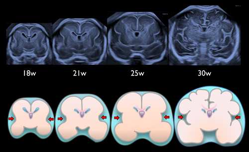
This figure was published by Dr. Pooh in an international journal in 2019. As you can see, the fetal brain changes its shape with the number of weeks, and CRIFM's Fetal brain dock shows how the brain changes with each week, along with various measurements.
Japan's first "Fetal Brain Center" established
Dr. Pooh has been observing the fetal brain for 25 years since 1996 and is internationally recognized for her work. She has contributed to academic journals and many English-language books on the fetal brain, and Dr. Pooh herself has published three books on the fetal brain.
Dr. Pooh, who has been in contact with many unborn babies with brain diseases and their siblings with various disabilities, has set up a dedicated floor named "Fetal Brain Center" on the fourth floor of the building at the end of 2019.
The spaciousness of the room was designed so that the disabled child could be taken in a wheelchair and mother could receive the Fetal brain dock in comfort.
Dr. Pooh was the first person to establish a fetal brain center in Japan, and the only one in the world. With 25 years of experience and expertise, we offer fetal brain dock for mid-term fetal Dock and late stage fetal Dock to all mothers. If a brain abnormality is suspected, the patient may proceed to counseling, and can opt genetic testing by Amniocentesis.
Examination at the Fetal Brain Center
What is fetal Neurosonography (brain scan)?
The Fetal brain dock was first developed by Dr. Pooh in 1996, when she began using transvaginal ultrasound to view the inside of the fetal brain.
In Japan, Dr. Pooh is a pioneer in this field, and the CRIFM fetal brain dock is famous overseas, and many doctors are interested in training at CRIFM.
Dr. Pooh closely observes the brain development of the fetus every day, so she can tell the number of weeks of pregnancy just by looking at the baby's brain.
In a normal prenatal checkup, the brain is simply sliced in a circle and the size of the head is simply measured. In CRIFM's Fetal brain dock, the brain is observed in various cross sections and various parts of the brain are measured in an original way, as shown in these figures, to check whether the fetal brain is developing normally.
Fetal brain dock is performed on all fetuses of patients who come to the hospital for mid-term fetal Dock and late stage fetal Dock.
However, depending on the position of the fetal head, we may need to take a break or ask the mother to walk around and wait for the fetus to change its position, so we recommend that you come to the hospital with plenty of time to spare.
Also, although there are individual differences in development, we recommend that you have a brain follow-up scan after one or two weeks if you have any concerns.
If you suspect that you have a genetic mutation, we will provide you with proper counseling, and if you wish to have a test, we will try to provide you with the results as soon as possible.
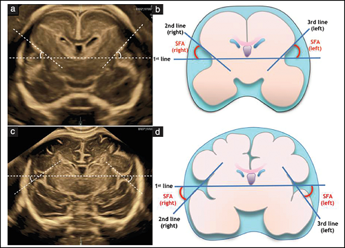
Measurement of the sylvian fissure angle in the fetal brain (an important factor in predicting future cortical development)
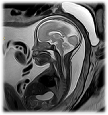
What is a fetal MRI scan?
CRIFM conducts a fetal neurological examination (brain dock) for diagnostic imaging, and an MRI scan can be used to complement the ultrasound scan to support the brain dock imaging diagnosis.
It is also advantageous as a means of imaging in cases where ultrasound imaging is difficult.
At CRIFM, in collaboration with the Umeda Brain, Spinal Cord and Neurology Clinic, MRI experts will perform fetal brain MRI imaging using the latest MRI technology (3.0 Tesla).
An MRI scan uses a magnetic field (the force of a magnet) and radio waves to image and capture information inside the body, so it does not use X-rays (radiation) like an X-ray or CT scan, and there is no exposure to radiation.
In addition, MRI scans are reported to have no adverse effects on the fetus, making it a very safe test.
Fetal brain MRI imaging with CRIFM is performed in the following cases.
- Unsuitable for neurosonography due to fetal position, gestational week, abnormal placental position, uterine fibroid, etc.
- Suspected fetal brain and nervous system abnormalities
- History of delivery of a child with neurological or developmental abnormalities
At the Umeda Brain, Spinal Cord and Neurology Clinic, the MRI imaging time for CRIFM is every Thursday from 12:20 to 14:20. However, in case of urgent cases, please consult with us.
What is a genetic test related to the brain development?
At CRIFM, we conduct a fetal neurological examination (brain dock) for diagnostic imaging. More than 100 genetic mutations related to fetal hydrocephalus and more than 100 genetic mutations related to cortical growth defects (MCD) have been reported.
Many of these genes are included in the Whole Exome Sequencing that we do at CRIFM, which can test for 6,704 genetic mutations.
In cases where fetal brain abnormalities are suspected, especially congenital abnormalities caused by genetic mutations, fetal genetic testing can be done using DNA from the chorionic tissue and amniotic fluid. In such cases, DNA from both parents is also required.
Congenital cerebral abnormalities caused by genetic mutations are also predicted to have a rather grim neurological prognosis after birth.
Of course, not all abnormalities can be explained by genetic mutations alone. Even if the results of the gene test are normal, the diagnosis will be made based on a comprehensive judgment that includes the status of the brain dock and brain follow-up scan.
In some cases, the genetic mutation is not detected in the amniotic fluid test, but the brain abnormality is caused by the increase of cells with the genetic mutation in a part of the brain.
Small, tiny genetic mutations can disrupt one of the steps in brain development and lead to severe disabilities.
Not everyone can take a genetic test. There are certain conditions that must be met before a genetic test can be done. In the field of neurology, it is limited to cases where a fetal neuro-ultrasound (brain dock) test has determined that there is a possibility of genetic mutation.
Fetal brain dock reveals various brain diseases
Fetal brain dock fee
Fetal brain dock is included in fetal Dock. The following are the fees for fetal Dock.
| Basic examination (per fetus) |
47,300 yen (tax included) *If you have already undergone the Mid-Trimester Fetal Screening, the basic fee for the Late-Trimester Fetal Screening is 27,500 yen (tax included). |
|---|---|
| Follow-up ultrasonography of the fetal brain | 14,300 yen and up (tax included) |
| Additional Detailed examination fee (per fetus) |
5,500 - 44,000 yen (tax included) *After basic inspection, add to basic fee if necessary |
| Additional Fetal Brain Quantitative Analysis fee (per fetus) |
11,000 - 27,500 yen (tax included) *After basic inspection, add to basic fee if necessary |
| Results counseling | 5,500 - 44,000 yen (tax included) |
About brain diseases in babies found in Fetal brain dock
1. Types of skull diseases
The fetus may have a skull that does not form properly for some reason. An acrania is when the entire skull fails to form because the neural tube fails to close properly, and the brain juts out of the skull.
There is also another disease called early craniosynostosis, in which the head and face gradually deform during pregnancy due to the fusion of the skull during fetal life, which is earlier than it should be. Early craniosynostosis is a disease caused by several genetic mutations, which can lead to facial features (exophthalmos, prominent forehead) and complications such as syndactyly and giant toes.
2. Early development of the cerebrum
The cerebrum separates into the left and right hemispheres at the very beginning of pregnancy, and the separation can be clearly seen on ultrasound from about 8 weeks of pregnancy.
If the cerebrum does not separate properly, it is called holoprosencephaly. Depending on the degree of separation, there are three types: alober, semilober and lober types.
The middle part of the face is often associated with dysplasia such as cleft lip and palate, arrhinia, elephant nose (proboscis), cyclopia or orbital narrowing.
3. Enlarged ventricles and hydrocephalus
Hydrocephalus is a condition in which cerebrospinal fluid is held back and does not flow out because of the obstruction of fluid flow pathway, and remains in the ventricles, resulting in enlargement of the ventricles.
In a similar situation, if the cerebrospinal fluid is flowing but the ventricles are large, it is not called hydrocephalus. In some cases, the ventricles are large because the cerebrum itself is developing slowly, which means that the cerebrum is thin compared to the number of weeks it has been in existence.
Hydrocephalus and enlarged ventricles are terms that describe a situation, not a disease. Whether it is hydrocephalus or enlarged ventricles, the cause must be clearly identified. The name of the disease varies depending on the cause.
In CRIFM, the cause is narrowed down based on the shape of the enlarged ventricles. It is also said that there are more than 100 gene mutations related to ventricular enlargement and hydrocephalus.
Once the genes responsible for ventricular enlargement and hydrocephalus are identified, it will be possible to predict the prognosis and degree of disability after birth.
4. Abnormal development of the corpus callosum
The nerve fibers that connect the right brain to the left brain are called the corpus callosum. When the corpus callosum does not form, it is called a corpus callosum agenesis.
If you have a cerebral corpus callosum agenesis, you need to be examined closely to see if there are any abnormalities in other parts of the brain or if there are any other complications. In some cases, it is caused by a genetic mutation, and in such cases, neurological symptoms are common.
If there is no other abnormality other than a defect in the corpus callosum and no genetic mutation is found, most babies will grow up healthy and without any symptoms at all.
It is also common for interhemispheric cysts (water balloons) to form between the right and left hemispheres of the brain, but since these balloons shrink and grow naturally, they are monitored periodically to determine if surgery is indicated after birth.
Lipomas can also form in the area of the corpus callosum, but these are benign and are not an indication for surgery.
5.Abnormal cortical development (MCD)
Malformations of cortical development (MCD) are closely related to brain development during the first half of pregnancy. Malformations of cortical development (MCD) have been shown to be caused by various genetic mutations.
Fetuses with MCD have poorly formed brain wrinkles (Lissencephaly, Pachygyria) or too many fine wrinkles (Polymicrogyria), which can lead to epilepsy and developmental disabilities, a condition that is often a problem in pediatric neurology.
There are quite a few different types, but they are broadly categorized into three types.
- Excessive cell growth or disorders in regulating the number of cells (megalencephaly, unilateral megalencephaly, cerebellopathy, etc.)
- Disorders when nerve cells move through the brain to create the cerebral cortex (such as Lissencephaly)
- Disorders of processes such as myelination after nerve cell migration (polymicrogyria)
6. Abnormalities of the cerebellum and posterior cranial fossa
In babies with spinal meningocele, a "Chiari II malformation" occurs. This is a disease in which the cerebellum and medulla oblongata are pulled down into the spinal canal, thus stopping the flow of spinal fluid and causing hydrocephalus.
Although it is called Chiari Type II malformation, it is not actually a malformation, nor is it a disease of the cerebellum itself.
Dandy-Walker malformation, which presents with partial or complete loss of the cerebellar appendages and cysts in the median posterior cranial fossa, and complications such as hydrocephalus, is known by name but is rare.
Very few babies referred to CRIFM with suspected Dandy-Walker malformation really have Dandy-Walker Syndrome. Rarely, babies with Dandy-Walker syndrome come to us, but since it is often associated with chromosomal abnormalities and genetic mutations, genetic testing is always necessary.
Joubert syndrome is not a brain disease, but a systemic disease. Joubert syndrome is a disease of the cilia (hair-like structures on many cells in the body that allow fluid to flow in the right direction) throughout the body.
In the brain, the ultrasound or MRI image looks like a slit in the brainstem with no cerebellar vermis, and is known as the "molar tooth sign".
There are currently 37 subtypes of Joubert's syndrome, each involving a different genetic mutation. Many children have a very severe form of the disease. Joubert syndrome is caused by a familial genetic mutation, which can be confirmed early in pregnancy with a CVS if the causative gene is known.
A disease in which the cerebellum does not divide properly into left and right parts, but clumps together, is called "rhombencephalosynapsis" and is said to be rare. However, it can go unnoticed at birth, and the true frequency of its occurrence is not known.
Some babies have hydrocephalus, which is a serious complication of the disease, and some children grow up healthy and large. The related gene for this disease has not yet been discovered.
7. Others
An arachnoid cyst is a cyst (water balloon) that develops in various places inside the skull and presses on the brain. They may tend to disappear during pregnancy, or they may grow in size. After the baby is born, the pediatric neurosurgery department will consider whether to operate on it.
A Vein of Galen aneurysmal malformation (VGAM) is a mass of blood vessels in the brain caused by a congenital abnormality of the blood vessels. Although it is called varicose veins, it is actually an arteriovenous shunt (a condition in which arteries and veins are connected without passing through peripheral blood vessels).
There are treatment options for VGAM, but it requires periodic detailed brain checks with neurosurgical ultrasound, as brain damage can occur frequently during fetal life due to circulation problems in the brain.
Other abnormalities that can be found during a fetal brain scan include the following.
There are various types of brain tumors, and it is necessary to diagnose whether they are malignant or benign, and also to determine the degree of bleeding associated with the tumor.
In cases of intracerebral hemorrhage, genetic mutations that predispose to hemorrhage may be found, and maternal platelet antibodies may also play a role. Depending on the situation, it is necessary to decide on the method and timing of delivery.
CRIFM is repeating brain scans in cooperation with delivery facilities in cases of intracerebral hemorrhage.
In some cases, calcified lesions in the brain are completely benign, but in other cases, the mother's infection (cytomegalovirus or toxoplasma) can be transferred to the fetus and cause multiple calcified lesions in the brain, requiring testing for cytomegalovirus antibodies.
Fetal brain outpatient care consultation process
Reservation
All consultations are by appointment only, and due to the large number of people who come for fetal Dock, we recommend that those who are concerned make an appointment as early as possible. Please make a reservation by phone or using the reservation form.
Please remember to bring your letter of introduction, health insurance card, and maternal and child health handbook with you when you come to the clinic. If you do not have a letter of introduction, an additional fee of 5,500 yen (including tax) will be charged.
In principle, mothers and fathers should visit the clinic together.
Reception desk
Please allow plenty of time for your visit, as there is always a wait. After the reception, the mother's blood pressure and weight will be measured.
medical examination
We take our time to examine the baby with ultrasound.
During the examination, mothers and fathers can also check the condition of the fetus on the monitor image.